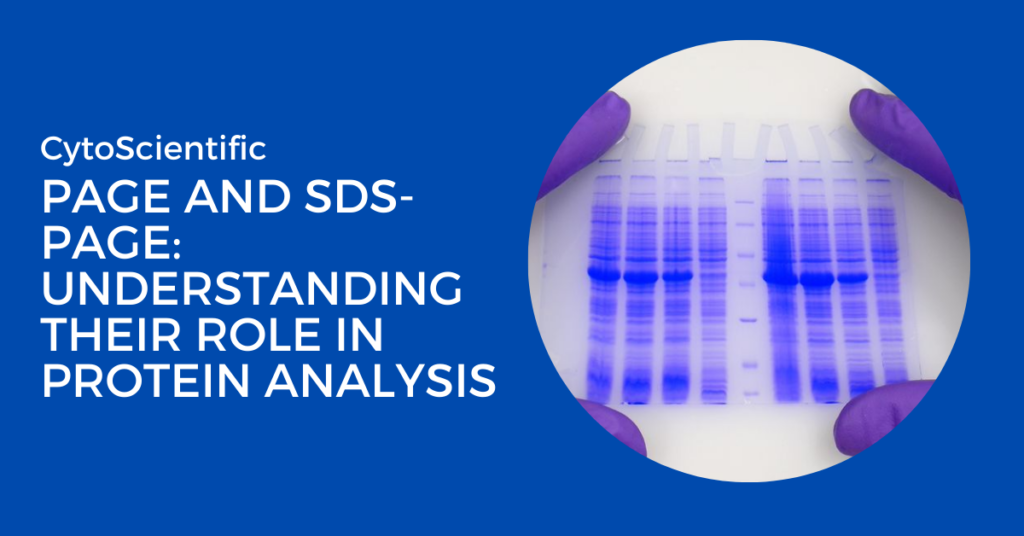
Two popular laboratory methods for separating substances such as proteins and nucleic acids are polyacrylamide gel electrophoresis (PAGE) and sodium dodecyl sulfate polyacrylamide gel electrophoresis (SDS-PAGE). They are essential to biochemistry, molecular biology, and other scientific fields.
PAGE and SDS-PAGE
PAGE divides molecules according to their charge, size, and shape. The method includes passing an electric current through a gel composed of polyacrylamide, a material that forms a structure like a mesh. Depending on their properties, the molecules are separated into different bands as they pass through the gel. PAGE is frequently used to separate nucleic acids or to investigate compounds in their native state.
Importance in protein analysis
How Does PAGE Work?
Polyacrylamide Gel Electrophoresis (PAGE) separates proteins or nucleic acids according to size, charge, and shape. Polymerizing polyacrylamide creates a porous matrix, which is then formed into a gel. Samples are placed in wells, and an electric field is applied. Molecules migrate across the gel towards the opposing charge, with smaller molecules moving more quickly. In native PAGE, proteins retain their inherent structure and charge, which influences their mobility. After running the gel, the molecules are seen with stains such as Coomassie Brilliant Blue, and the migration pattern is used to estimate their size and purity. PAGE is commonly employed in protein analysis and study.
Types of PAGE
Native PAGE
Native PAGE separates proteins or nucleic acids without denaturing them. This means that the molecules retain their normal structure, charge, and biological activity
Denaturing PAGE
In this technique, chemicals like as urea are employed to break secondary structures, separating molecules based on size under denatured circumstances.
How SDS-PAGE Separates Proteins
SDS-PAGE (Sodium Dodecyl Sulfate Polyacrylamide Gel Electrophoresis) separates proteins based on size. SDS, a detergent, is used to denature proteins, giving them a uniform negative charge and unfolding their structure. This eliminates the effect of protein shape or charge, so separation depends solely on size. Samples are loaded into a polyacrylamide gel, and an electric field is applied, causing proteins to migrate towards the positive electrode. Smaller proteins travel faster through the gel. After the run, the proteins are stained, and their size is determined by comparing migration to a molecular weight marker. SDS-PAGE is key in protein analysis.
Key Components of PAGE and SDS-PAGE
Both techniques require essential components for successful electrophoresis:
- Buffers: Maintain pH stability and ion strength.
- Gel Casting Tools: Ensure uniform gel preparation.
- Electrophoresis Apparatus: Facilitates controlled movement of molecules.
Comparison Between PAGE and SDS-PAGE
| Feature | PAGE | SDS-PAGE |
|---|---|---|
| Separation Principle | Separates based on size, charge, and shape. | Separates based on size only after denaturation. |
| Protein Structure | Proteins retain their natural, folded state. | Proteins are denatured and unfold. |
| Effect of SDS | No SDS is used, so proteins are in their native state. | SDS is used to denature proteins and give them a uniform negative charge. |
| Migration Pattern | Migration depends on both size and charge. | Migration is based solely on size. |
| Applications | Studying protein complexes, enzyme activity, and protein interactions. | Determining protein molecular weight, analyzing protein purity, and studying complex mixtures. |
| Sample Preparation | Proteins are not altered; minimal preparation needed. | Requires treatment with SDS and usually a reducing agent to break disulfide bonds. |
| Gel Composition | Polyacrylamide gel with a single concentration for resolving proteins. | Polyacrylamide gel can be used with a gradient concentration for better resolution. |
| Results | Provides insights into protein function and interactions. | Provides molecular weight estimation and purity assessment. |
| Type of Proteins | Suitable for analyzing proteins in their native state, preserving biological activity. | Best for analyzing denatured proteins where size is the primary focus. |
| Visualization | Bands represent native protein forms and their complexes. | Bands represent denatured proteins, useful for size comparison. |
Conclusion
While both PAGE and SDS-PAGE are valuable tools in protein analysis, they serve different purposes. PAGE is ideal when it is essential to preserve the natural state and functionality of proteins, making it suitable for studying protein interactions and complexes. SDS-PAGE, on the other hand, is primarily used when the focus is on the molecular weight and purity of proteins, as it denatures proteins and standardizes their charge for size-based separation. Both techniques are essential in molecular biology, biochemistry, and proteomics for understanding the characteristics and behavior of proteins.

